Advancing Prehospital Care with Point-of-Care Ultrasound (POCUS): Enhancing Diagnosis and Decision-Making in the Field
Aicha M. Hull, MD, AEMUS-FPD
Madigan Army Medical Center
Hillary Harper, MD, AEMUS-FPD, FACEP
Madigan Army Medical Center
Anders B. Conway, MD
Thurston County Medic One
Mitch Arends, MD
Madigan Army Medical Center
Myles Melton, MD
Providence St. Peter Hospital
Introduction
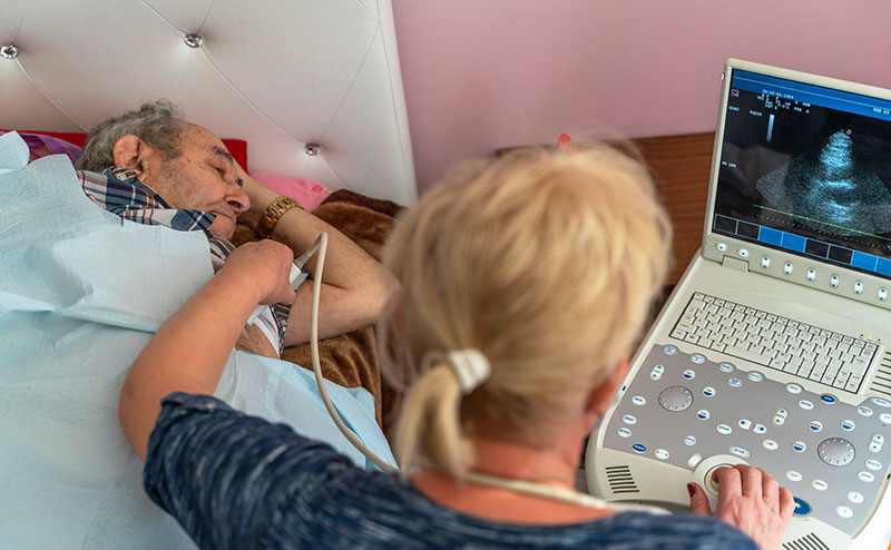
Point-of-care ultrasound (POCUS) has become an indispensable tool for prehospital providers—paramedics, emergency medical technicians (EMTs), and flight medics—enabling rapid assessment and diagnosis of critically ill or injured patients in the field.1 Real-time, on-site imaging allows for quicker and more accurate diagnoses, supports treatment decisions, and facilitates timely interventions, all of which can improve patient outcomes even before reaching the hospital. With proper training and careful device selection, prehospital providers can seamlessly integrate ultrasound into their practice, bolstering their ability to deliver critical care during emergency situations. As technology advances and ultrasound devices become increasingly portable and accessible, the role of POCUS in prehospital medicine is set to expand significantly.
Clinical Applications in Prehospital Settings
In the prehospital environment, POCUS serves a variety of critical functions. This includes trauma care, out-of-hospital cardiac arrest (OHCA), shock and respiratory distress. The Enhanced Focused Assessment with Sonography in Trauma (eFAST) exam allows providers to quickly assess for internal bleeding in the abdomen, thoracic cavity, or pericardium.2 Identifying life-threatening conditions such as hemoperitoneum, pneumothorax, or cardiac tamponade can prompt urgent interventions such as life-flight transport and activation of trauma teams at the receiving hospital.
In cases of cardiac arrest, POCUS can be used to detect organized cardiac motion that may not be visible during a simple pulse check in a severely hypotensive patient. Ultrasound can also inform the team when continued resuscitation efforts may be futile, such as when cardiac standstill is observed after prolonged efforts.3
Another essential application is in the assessment of shock. Basic techniques, like evaluating the diameter and collapsibility of the inferior vena cava (IVC), provide insight into a patient’s volume status and guide fluid resuscitation. More advanced protocols, such as the Rapid Ultrasound in Shock and Hypotension (RUSH) exam can rule out life-threatening conditions such as pericardial tamponade or abdominal aortic aneurysm (AAA). POCUS can also be used to detect new-onset acute heart failure through limited echocardiography and identification of lung pathology such as multiple B-lines on thoracic ultrasound, indicating the need for immediate BiPAP intervention.4
Improved Patient Management
The integration of POCUS into prehospital care significantly enhances decision-making. With real-time diagnostic capabilities, paramedics and flight medics can confirm conditions like pneumothorax or hemothorax in trauma patients. This can prevent both delays in care as well as help avoid unnecessary procedures. Prehospital protocols that incorporate airway management applications can also help rapidly detect endotracheal tube dislodgement or confirm effective tube placement, improving patient safety and outcomes.5
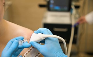
While urban settings with rapid transport times may see less frequent use of musculoskeletal assessments, POCUS can be especially valuable in rural settings. It allows for real-time evaluation of fractures, dislocations, joint effusions, and tendon tears, which can supplement physical examination and offer a more detailed assessment.6 This is particularly useful in identifying the location and nature of injuries, helping guide immediate on-site treatment or decisions during transport.
Training and Limitations
As with all advanced medical technologies, proper training is essential to maximize the benefits of POCUS.7 Although ultrasound devices have become more portable and user-friendly, they still require a solid understanding of ultrasound physics, probe selection, and image interpretation. Prehospital providers must undergo specialized training in basic ultrasound techniques - focusing on core applications such as trauma, OHCA, shock, respiratory distress, and perhaps musculoskeletal injuries.
Despite these advancements several challenges remain, particularly related to image quality in the field. Environmental factors like lighting, patient positioning, background movement and device limitations can impact the accuracy of ultrasound exams. The skills required to perform high-quality scans and interpret images correctly demands continuous practice and ongoing learning.8 Providers must also be able to recognize when ultrasound findings are inconclusive and know when to seek further evaluation upon arrival at the hospital.
Additionally, there is a risk of misinterpretation in certain contexts. For example: while a hospital provider might recognize a dilated gallbladder as a normal anatomical feature, a prehospital provider could mistakenly identify it as free fluid in the right upper quadrant and thus a false positive. This underscores the importance of familiarity with normal anatomic variants and the limitations of ultrasound in prehospital settings.
Conclusion
POCUS has proven to be a game-changer in prehospital care - allowing providers to make faster, more accurate clinical decisions and significantly improving patient outcomes in the field. With continued training, appropriate device selection, and thoughtful integration into prehospital protocols, providers can use ultrasound to enhance critical care delivery in emergency situations. As ultrasound technology evolves and becomes even more accessible and portable, the role of POCUS in prehospital medicine will undoubtedly continue to grow, offering increased opportunities for life-saving interventions. However, the successful implementation of prehospital POCUS programs must address unique challenges, including training, device limitations, and the need for ongoing education and quality assurance to ensure accurate and effective use in the field.
Disclaimer
The views expressed are those of the author(s) and do not reflect the official policy of the Department of the Army, the Department of Defense, or the U.S. Government.
References
- Vianen NJ, Van Lieshout EMM, Vlasveld KHA, Maissan IM, Gerritsen PC, Den Hartog D, Verhofstad MHJ, Van Vledder MG. Impact of Point-of-Care Ultrasound on Prehospital Decision Making by HEMS Physicians in Critically Ill and Injured Patients: A Prospective Cohort Study. Prehosp Disaster Med. 2023;38(4):444-9.
- Gharahbaghian L, Anderson KL, Lobo V, Huang RW, Poffenberger CM, Nguyen PD. Point-of-Care Ultrasound in Austere Environments: A Complete Review of Its Utilization, Pitfalls, and Technique for Common Applications in Austere Settings. Emerg Med Clin North Am. 2017;35(2):409-41.
- Hussein L, Rehman MA, Sajid R, Annajjar F, Al-Janabi T. Bedside ultrasound in cardiac standstill: a clinical review. Ultrasound J. 2019;11(1):35.
- Russell FM, Supples M, Tamhankar O, Liao M, Finnegan P. Prehospital lung ultrasound in acute heart failure: Impact on diagnosis and treatment. Acad Emerg Med. 2024;31(1):42-8.
- Weaver B, Lyon M, Blaivas M. Confirmation of endotracheal tube placement after intubation using the ultrasound sliding lung sign. Acad Emerg Med. 2006;13(3):239-44.
- Anderson A, Theophanous RG. Point-of-care ultrasound use in austere environments: A scoping review. PLoS One. 2024;19(12):e0312017.
- Al-Absi DT, Simsekler MCE, Omar MA, Soliman-Aboumarie H, Abou Khater N, Mehmood T, Anwar S, Kashiwagi DT. Evaluation of point-of-care ultrasound training among healthcare providers: a pilot study. Ultrasound J. 2024;16(1):12.
- Pinto A, Pinto F, Faggian A, Rubini G, Caranci F, Macarini L, et al. Sources of error in emergency ultrasonography. Crit Ultrasound J. 2013;5 Suppl 1(Suppl 1):S1.


