Cracking the Case: Rib Fractures
Contributing Author:
Huy Alex Duong, MD
Emergency Medicine Resident, UCLA Ronald Reagan/Olive View
Case
A 50-year-old male presents to the Emergency Department for right sided chest pain after slipping down a set of bleachers while doing his routine morning stair climbing. He fell directly onto the right side of his chest, did not lose consciousness, but now reports severe pain that has been unrelieved with NSAIDs at home. His vital signs are reassuring without any signs of hypoxia. By the time the patient presents, it is noon and the department is getting busy. Your leading diagnosis is traumatic rib fractures and as you reach to click for the X-ray order, you wonder if you need to consider any other diagnostic tools.
X-ray
X-rays are typical first line imaging studies obtained when a patient presents with blunt chest injury as they are low-radiation risk, relatively inexpensive, and quick to obtain. In addition to identifying obvious displaced rib fractures, they are also used to identify more severe complications of rib fractures such as pneumothoraces, hemothoraces, flail chest, or pulmonary contusions. Feared complications including pneumonias in elderly patients may be picked up on X-ray as late-stage findings though negative x-rays do not exclude its diagnosis1. Though often ordered, X-rays are notoriously poor for appropriate identification of rib fractures and some studies have identified up to 50-75% of rib fractures missed on X-ray imaging are later identified on CT chest2. It is more important to identify clinically significant rib fractures such as those with the associated complications listed above, multiple rib fractures (>3) especially in elderly patients with cardiopulmonary disease, or major displacement as these lend to worsening morbidity and mortality2. Most isolated rib fractures not identified on X-ray imaging go on to be discharged with appropriate pain control and pulmonary rehabilitation. Though the risk of mortality is low, studies have suggested long-term functional deficits3.
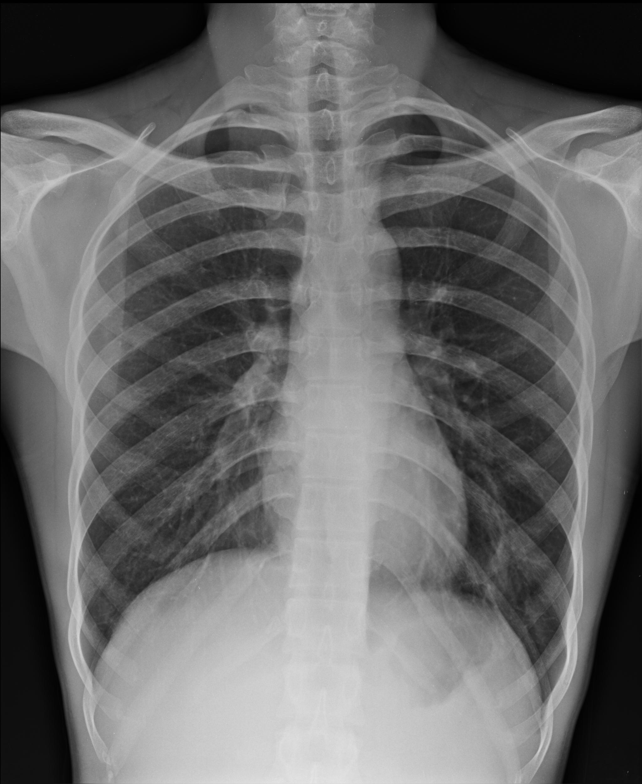
Figure 1:
PA chest x-ray.
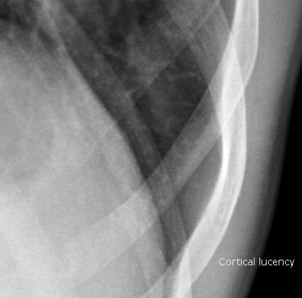
Figure 2:
Magnified image showing subtle cortical lucency of the lower rib suggesting fracture.
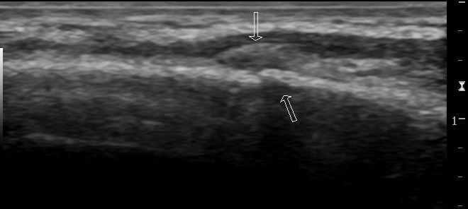
Figure 3:
Longitudinal view demonstrating a cortical break with overlying crescent shaped hematoma of the same rib fracture as figures 1 and 2.
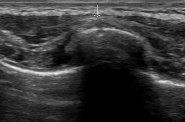
Figure 4:
Transverse view again demonstrating a cortical break with overlying hematoma.
The example images and annotations are courtesy of Dr. Maulik S Patel, Radiopaedia.org. From the case rID: 16479
Ultrasound
Point-of-care ultrasound (POCUS) is becoming increasingly common in emergency departments and its uses are expanding as they become more prevalent tools. Use of ultrasound has been shown in some studies to be more sensitive than chest x-rays for diagnosis of rib fractures4,5.
Using a linear probe, place it perpendicular to the rib obtaining a transverse view and tracing the length of the rib at the site of highest tenderness. Observe for cortical disruption and surrounding hematoma as this can suggest a rib fracture. A longitudinal view should also be obtained for confirmation of findings.
Although readily available, POCUS is highly dependent on the skill of the operator and can miss multiple rib fractures6,7. Utilization of ultrasound can be used in aiding diagnosis of rib fractures but like x-rays, clinical context is important in a patient’s ultimate disposition.
CT
CT chest imaging is regarded as the gold standard for diagnoses of suspected rib fractures and most studies compare CT imaging with other diagnostic modalities such as those listed above. Though more sensitive in diagnosis, its clinical utility is questionable in uncomplicated rib fractures, and CT is typically more costly, time consuming, and carries a higher risk of radiation without changing clinical management4.
Identifying factors that influence prognosis is essential. Increasing number of rib fractures, advanced age, major displacement, and presence of associated injuries (i.e. hemothorax, pneumothorax, aortic injury, or flail chest) can all contribute to higher risks of mortality and morbidity. In these situations, should your clinical suspicion be high or the mechanism of injury is concerning, comprehensive CT imaging, possibly extending beyond the chest, is often justified to assess for extent of injury.
Case Conclusion
The patient ultimately received a 2-view PA/lateral chest X-ray which revealed 2 acute rib fractures on the right side. His vitals remained stable without hypoxia and his pain was well controlled in the Emergency Department with opioids. He was ultimately discharged home without further workup with a prescription for lidocaine patches, methocarbamol, and oral opioids for breakthrough pain. He was also given an incentive spirometer for pulmonary rehabilitation. On 1-month follow up, his pain is improving, and he has not had any new fevers, shortness of breath, or any other symptoms.
References
- Self WH, Courtney DM, McNaughton CD, Wunderink RG, Kline JA. High discordance of chest x-ray and computed tomography for detection of pulmonary opacities in ED patients: implications for diagnosing pneumonia. Am J Emerg Med. 2013;31(2):401-405. doi:10.1016/j.ajem.2012.08.041
- Chapman BC, Overbey DM, Tesfalidet F, et al. Clinical Utility of Chest Computed Tomography in Patients with Rib Fractures CT Chest and Rib Fractures. Arch Trauma Res. 2016;5(4):e37070. Published 2016 Sep 13. doi:10.5812/atr.37070
- Émond M, Sirois MJ, Guimont C, et al. Functional Impact of a Minor Thoracic Injury: An Investigation of Age, Delayed Hemothorax, and Rib Fracture Effects. Ann Surg. 2015;262(6):1115-1122. doi:10.1097/SLA.0000000000000952
- Expert Panel on Thoracic Imaging:, Henry TS, Donnelly EF, et al. ACR Appropriateness Criteria® Rib Fractures. J Am Coll Radiol. 2019;16(5S):S227-S234. doi:10.1016/j.jacr.2019.02.019
- Vassalou EE, Perysinakis I, Klontzas ME, de Bree E, Karantanas AH. Performance of thoracic ultrasonography compared with chest radiography for the detection of rib fractures using computed tomography as a reference standard. Skeletal Radiol. 2024;53(11):2367-2376. doi:10.1007/s00256-024-04658-8
- Çelik A, Akoglu H, Omercikoglu S, et al. The Diagnostic Accuracy of Ultrasonography for the Diagnosis of Rib Fractures in Patients Presenting to Emergency Department With Blunt Chest Trauma. J Emerg Med. 2021;60(1):90-97. doi:10.1016/j.jemermed.2020.06.063
- Battle C, Hayward S, Eggert S, Evans PA. Comparison of the use of lung ultrasound and chest radiography in the diagnosis of rib fractures: a systematic review. Emerg Med J. 2019;36(3):185-190. doi:10.1136/emermed-2017-207416