Saphenous Nerve Block
Leonard V. Bunting, MD, FACEP
I. Overview and Indications
- The saphenous nerve is a sensory only nerve and is the largest cutaneous branch of the femoral nerve.
- Anesthesia of the medial lower leg can be achieved by blocking either the femoral or saphenous nerves.
- The saphenous nerve can be blocked anywhere along its course, from its origin under the sartorius muscle to its terminal branches near the ankle.
- This section will cover blockade of the nerve below the knee, after its association with the saphenous vein.
Anatomy
- The saphenous nerve divides off the femoral nerve in the proximal thigh
- It follows the superficial femoral vessels in the medial thigh, deep to the sartorius muscle.
- Proximal to the knee the nerve emerges superficially to associate with the greater saphenous vein.
- The nerve then continues down the medial lower leg with the greater saphenous vein to lie anterior to the medial malleolus at the ankle.
- Blockade of the nerve by the knee anesthetizes the medial lower leg/foot and blocking at the ankle provides anesthesia to a small medial portion of the ankle and foot.
Indications
- Injuries to medial leg, including medial malleolus fractures
Contraindications
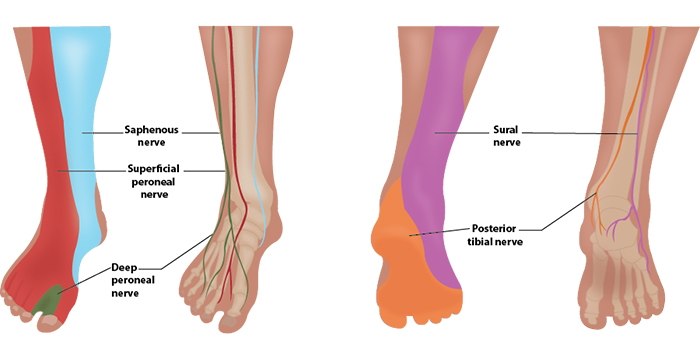
Illustration 1. Distribution of Anesthesia
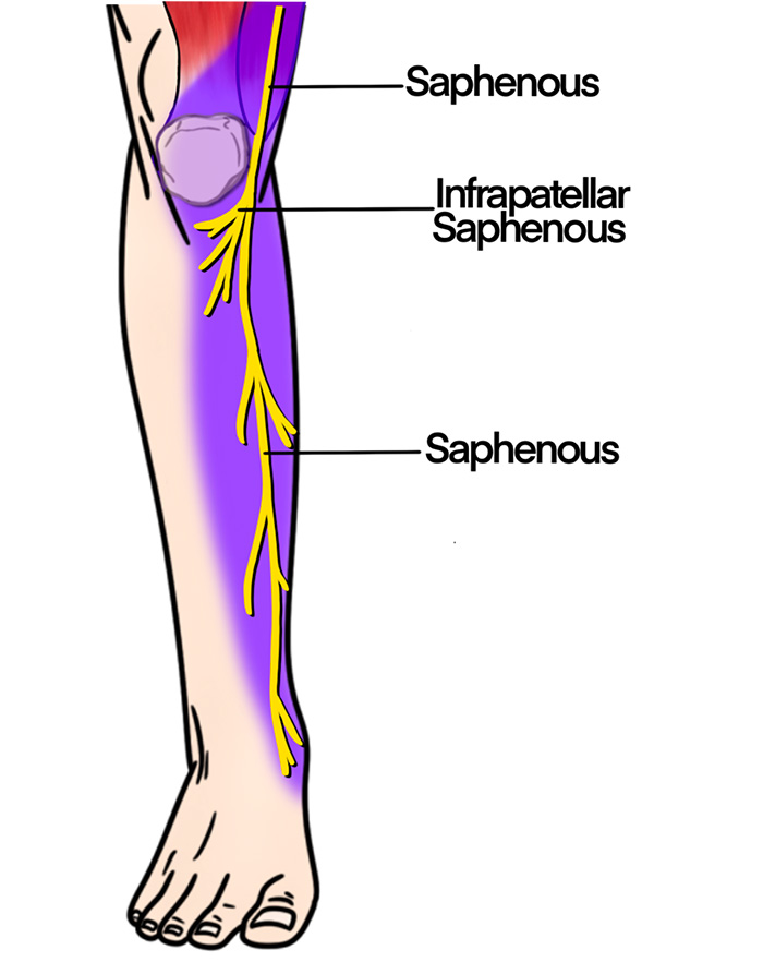
Illustration 2. Course of the saphenous nerve
II. Equipment
- Probe selection: 12-18 MHz linear transducer
- Sterile transparent film dressing (eg, Tegaderm™) or sterile probe cover
- 5ml of anesthetic
- 25-30-gauge needle for skin wheal with syringe of 2-3 ml of lidocaine with epi
- 22-25-gauge needle, 1.5 inch or longer (Needle choices) depending on body habitus
III. Setup and Patient Positioning
- General procedure setup
- Multiple positions can be used to expose the medial leg, such as externally rotating the leg and flexing the knee.
IV. Pre-scan/Sonographic Anatomy
- A high-frequency linear array probe is applied in a transverse plane anterior to the medial malleolus.
- Identify the greater saphenous vein anterior and superficial to the medial malleolus.
- The vein will collapse easily in most patients, so a tourniquet placed around the calf and light probe pressure are likely required.
- The small, echogenic saphenous nerve may be identified adjacent to the vein in any orientation, but it is commonly very difficult to see.
- If the nerve is not apparent, tracing the vein proximally may highlight the hyperechoic nerve following the vein.
- If the nerve is still not visualized, the saphenous vein is used as the target for injection.
- Trace the nerve and/or vein proximally to identify the optimal block location
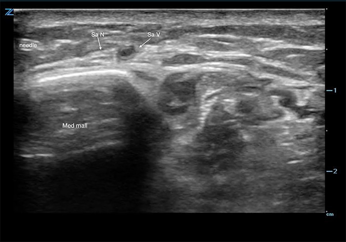
Figure 1. Saphenous nerve
Video 1. Pre-scan of the saphenous nerve
V. Procedure Technique
- General procedure setup
- Cover probe using sterile transparent film dressing (eg, Tegaderm™) or sterile probe cover.
- Flush block needle with a small amount of anesthetic to remove air.
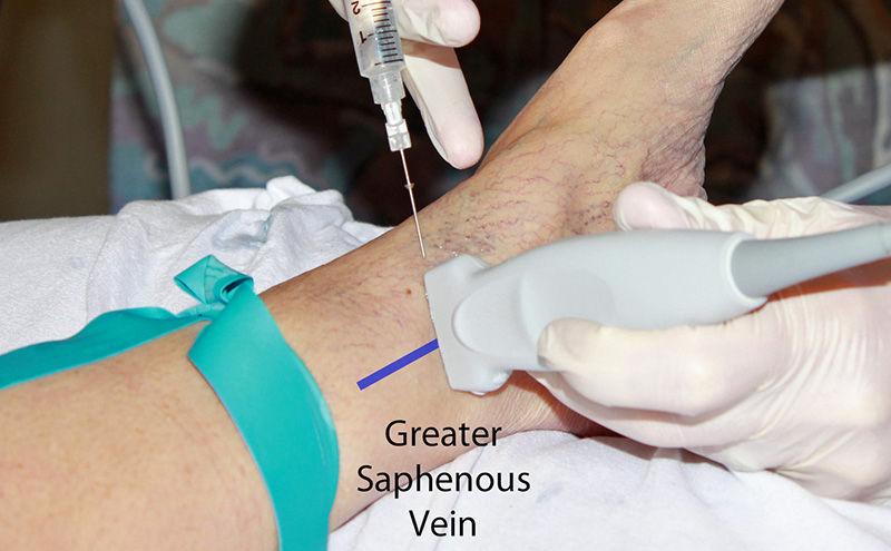
- An in-plane approach is preferred.
- Identify the nerve anywhere from the knee to the ankle, as detailed in the pre-scan section above.
- If nerve is not visible, deposit local anesthetic immediately anterior and posterior to the greater saphenous vein.
- The nerve will generally become visible during injection.
- After skin anesthesia, insert the block needle 3 mm at the short side of the probe at a shallow needle angle.
- Identify the needle tip by sliding the probe towards and then across the block needle (Visualizing the needle).
- Slowly advance the needle towards the deep, proximal border of the nerve (or greater saphenous vein).
- Once movement of the needle causes movement on the nerve (ie ‘mechanical coupling’), inject 0.5 cc of anesthetic.
- Follow injection precautions.
- If the anesthetic flows around the nerve, continue to inject in 1 cc increments until the nerve is surrounded and the block volume is reached.
- If the anesthetic is seen outside the perineural space, redirect the needle and inject another 0.5 cc.
- Readjustment of the needle position may be necessary to achieve adequate distribution of anesthesia.
- Always perform aspiration and incremental injection to avoid systemic distribution of the anesthetic.
- Typical block volumes are 3–5 cc.
- Full block onset may take up to 15–20 minutes, particularly if a long-acting anesthetic was used.
- Identify the nerve anywhere from the knee to the ankle, as detailed in the pre-scan section above.
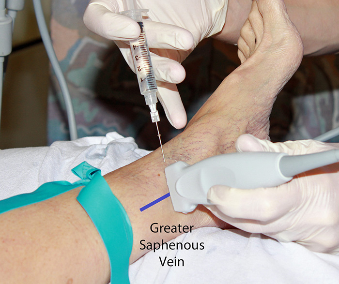
Figure 2. Saphenous nerve block in-plane hands
Video 2. Saphenous nerve block in-plane
VI. Post-procedure Care
- None required.
- Consider marking on skin with skin pen the time and date of block performed.
VII. Pearls and Pitfalls
- Clear all needles and syringes of air prior to injecting.
- The saphenous nerve is commonly not seen. Aim for the saphenous vein to start, and the nerve will likely become apparent during injection.
- Ensure spread of local anesthetic between the vein and nerve.
- Placing a rolled towel or pillow underneath the calf may aid in positioning for the procedure.
- Smaller needles are more difficult to visualize using ultrasound. Novices should consider using 23-gauge needles to start.
- Avoid vascular injection and injury by frequently aspirating. Any injected anesthetic should result in a bolus of fluid on the image. If no bolus is seen after injection, this may indicate intravascular injection.
- For patients with little subcutaneous tissue, the probe may not sit well near the medial malleolus. It is sometimes necessary to perform the procedure more proximal to avoid bony projections.


