Superficial Peroneal Nerve Block
Leonard V. Bunting, MD, FACEP
I. Overview and Indications
- The superficial peroneal nerve provides sensory innervation to a large portion of the anterior foot and therefore blocking it is very useful in the emergency setting.
- Although traditionally blocked with a field approach, ultrasound can be used to identify and block this nerve either at the ankle or more proximally.
- The superficial peroneal nerve is a mixed nerve that provides motor innervation to the fibularis and extensor digitorum longus muscles and cutaneous innervation to the anterior lateral ankle and dorsum of the foot.
Indications
- Injuries to the anterior foot, especially burns and lacerations
- Midfoot and forefoot fractures either alone or combined with tibial and/or sural nerve blocks
Contraindications
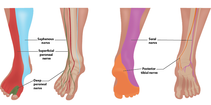
Illustration 1. Distribution of anesthesia
II. Equipment
- Probe selection: 12-18 MHz linear transducer
- Sterile transparent film dressing (eg, Tegaderm™) or sterile probe cover
- 5ml of anesthetic
- 25-30-gauge needle for skin wheal with syringe of 2-3 ml of lidocaine with epi
- 22-25-gauge needle, 1.5 inch or longer (Needle choices) depending on body habitus
III. Setup and Patient Positioning
- General procedure setup
- To expose the lateral leg and anterior foot the leg is internally rotated but multiple patient positions are possible.
IV. Pre-scan/Sonographic Anatomy
Anatomy
- The common peroneal nerve branches off the sciatic nerve above the popliteal fossa and courses laterally to the fibular head.
- At the proximal fibula, the common peroneal nerve divides into the deep peroneal and superficial peroneal nerves.
- The superficial peroneal nerve continues down the lateral aspect of the leg and innervates the peroneus muscles.
- In the mid lower leg, it runs in the superficial portion of a groove between the peroneus brevis and extensor digitorum longus muscles.
- In the distal lower leg, it forms superficial perforating branches that run over the lateral ankle and onto the anterolateral foot.
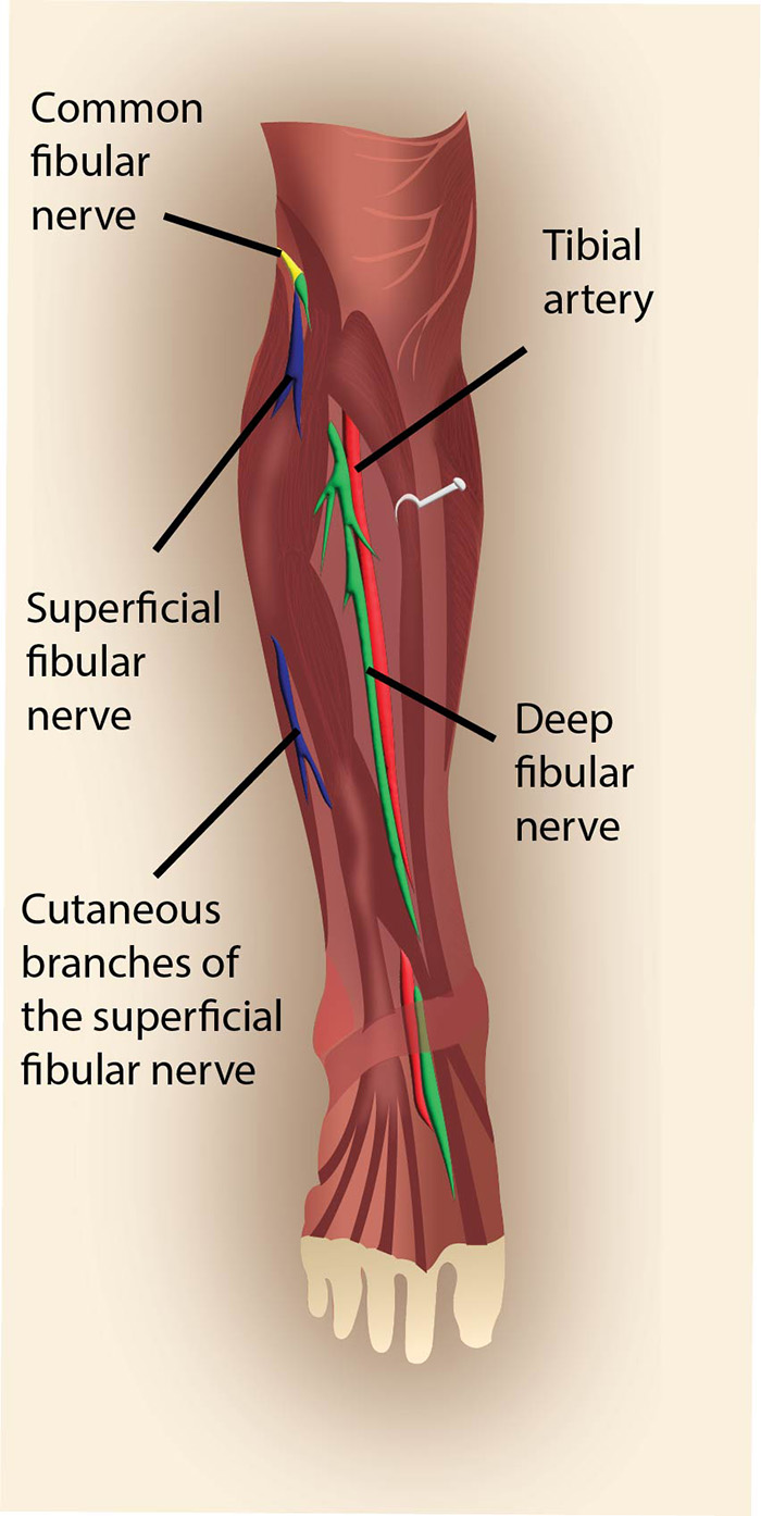
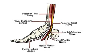
Illustration 2. Course of the superficial peroneal nerve
Video 1. Pre-scan of the superficial peroneal nerve
- The superficial peroneal nerve can be followed from its origin near the fibular head to its terminal branches on the dorsum on the foot.1
- A high frequency (12-15 MHz) probe is placed transversely on the lateral lower leg just above the lateral malleolus with the probe indicator pointed towards the operator's left.
- Identify the fibula as a hyperechoic arc with dense posterior shadowing.
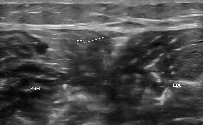
- Follow the fibula proximally to its midpoint where the peroneus brevis and extensor digitorum longus muscles are seen (Figure 1).
- These two muscles meet above the fibula.
- The superficial peroneal nerve travels in their superficial border and is seen as a hyperechoic oval or triangular shape.
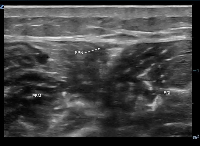
Figure 1. Superficial peroneal nerve, proximal
- Once identified, the nerve may be followed distally to the anterior ankle, where it appears as a subtle group of hypoechoic circles spread out in the very superficial cutaneous planes.
- Although the nerve can be blocked anywhere along its course, proximal sites can cause weakness and therefore may be less favorable in ambulatory patients.
V. Procedure Technique
- General procedure setup
- Cover probe using sterile transparent film dressing (eg, Tegaderm™) or sterile probe cover
- Flush block needle with small amount of anesthetic to remove air
- Superficial peroneal block approach
- An in-plane technique is preferred.
- Identify the nerve in the fascial plane between the fibularis and extensor digitorum longus muscles.
- Can be blocked anywhere along its course
- Distally near the ankle and foot the nerve can be hard to visualize with standard bedside probes. If seen it will appear as a hypoechoic bundle in the superficial planes.
- After skin anesthesia, insert the block needle 3 mm at the short side of the probe.
- Identify the needle tip by sliding the probe towards and then across the block needle. (Visualizing the needle)
- Slowly advance the needle towards the deep, proximal border of the nerve.
- Once movement of the needle causes movement on the nerve (ie ‘mechanical coupling’), inject 0.5 cc of anesthetic.
- Follow injection precautions.
- If the anesthetic flows around the nerve, continue to inject in 1 cc increments until the nerve is surrounded and the block volume is reached.
- If the anesthetic is seen outside the perineural space, redirect the needle and inject another 0.5 cc.
- Readjustment of the needle position may be necessary to achieve adequate distribution of anesthesia.
- Typical block volumes are 3–5 cc.
- Full block onset may take up to 15–20 minutes, particularly if a long-acting anesthetic was used.
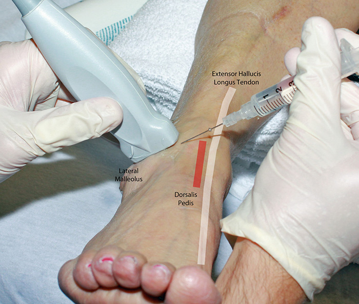
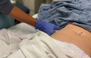
Figure 2. Superficial peroneal nerve block in-plane hands
Video 2. Superficial peroneal nerve block in-plane
VI. Post-procedure Care
- None required.
- Consider marking on skin with skin pen the time and date of block performed.
VII. Pearls and Pitfalls
- If the nerve is not readily apparent at the mid-fibula, anesthetic injected between the two muscle groups may aid in visualization.
- The superficial peroneal nerve is best seen at the ankle by tracing it from proximal. Attempting to identify it by starting at the ankle is challenging.
- Placing a rolled towel or pillow underneath the calf may aid in positioning for the procedure.
- Smaller needles are more difficult to visualize using ultrasound. Novices should consider using 22-gauge needles to start.
VIII. Reference
- Canella C, Demondion X, Guilin R, et al. Anatomic study of the superficial peroneal nerve using sonography. AJR Am J Roentgenol. 2009;193(1):174-9.


